Beyond Morphological Dilation: Revisiting Conformal Prediction for 3D Tumor Segmentation
- Uncertainty Quantification
- Computer Vision
We separate boundary errors from missed tumor components and combine component-aware calibration with confidence-guided geodesic expansion.
Paper UNSURE 2026
- EURECOM
A segmentation model can delineate the main tumor accurately while missing a disconnected focus. A margin calibrated by conformal prediction (CP) must therefore describe both boundary error and missed tumor tissue. We show how capped morphological dilation can certify its calibration score while leaving the requested tumor coverage unmet. Our solution separates these errors and expands along model-confidence paths. On fixed BraTS 2021 predictions, the combined construction removes the observed vacuity across three tumor regions and reports the missed-volume uncertainty explicitly.
Why a calibrated dilation can miss the tumor
Let be the input image, the predicted mask, the reference mask, and a nested family of expanded masks. The score measures the expansion needed to include a fraction of the tumor:
For target miscoverage , split conformal calibration takes the -th ordered score from exchangeable calibration cases, using when that order exceeds . For dilation, is a radius. The dilation baseline searches only up to voxels and caps scores when the requested coverage remains unreachable on that grid.
This cap changes the certified event. A test score can fall below the calibrated cap while the expanded mask still misses tumor tissue. We call the resulting discrepancy vacuity. Both distant missed foci and large boundary errors can cause saturation.

Boundary errors and missed components
For region , we match each reference component to the prediction when their overlap reaches voxels, with by default. Let contain the matched components and contain the unmatched tissue. Instance-level CP uses
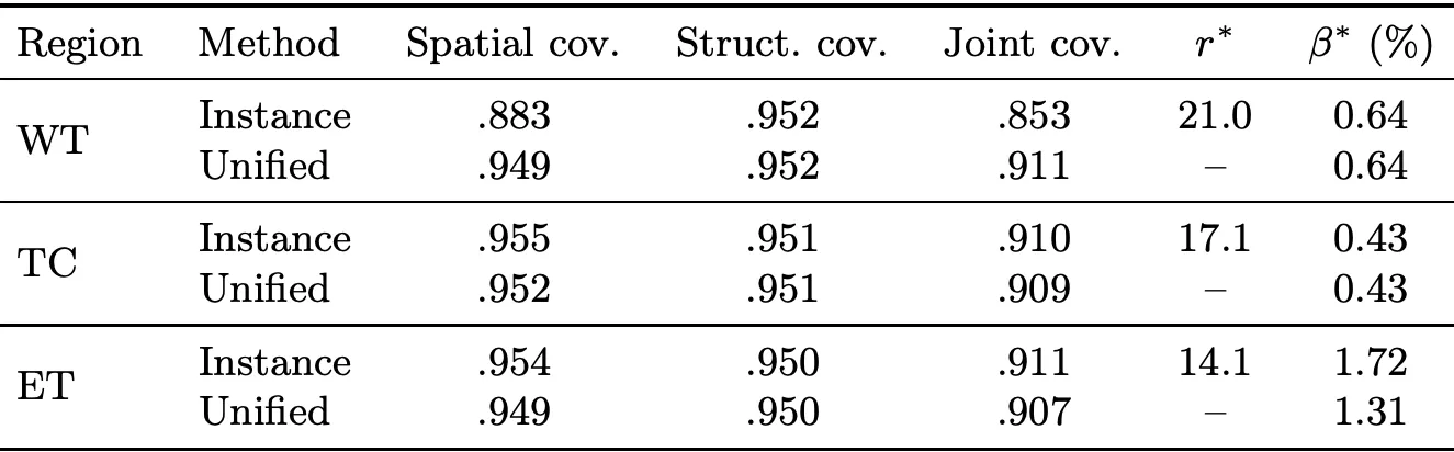
Calibrating each score at miscoverage gives a radius and missed-volume bound . Under exchangeability and with the spatial event represented by its score, a union bound gives
The spatial clause holds by convention when is empty. The certificate describes coverage of detected components and bounds the volume of missed tissue, whose location is unknown at prediction time. A capped spatial score can still saturate, motivating the geodesic construction.
Expansion along confidence-guided paths
We replace isotropic distance with an arrival-time field computed by Fast Marching. For voxel and region probability ,
Expansion travels faster through probable tumor tissue. The positive speed floor permits travel through low-confidence background and gives finite arrival times for disconnected foci. Geodesic CP calibrates the -quantile of over and returns . The unified method calibrates arrival times over matched components and retains the structural score.
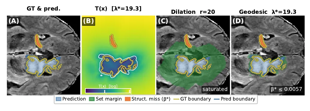
Evidence from fixed BraTS predictions
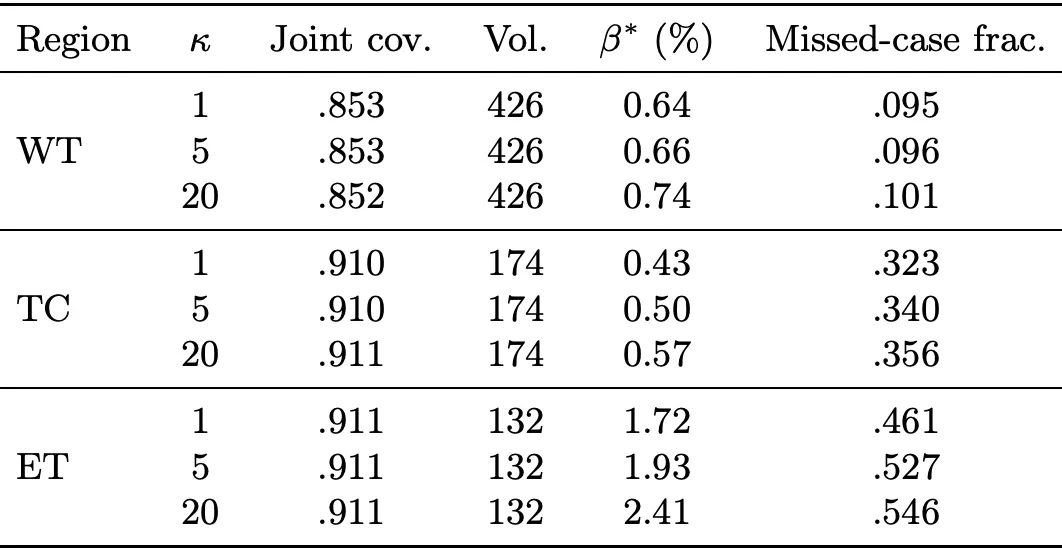
We retain 1,239 BraTS 2021 cases with complete predictions and probability maps from a fixed segmentation model. Each of 100 splits uses 991 calibration and 248 test cases, with . Reference components below 100 voxels are removed before scoring. Whole tumor (WT), tumor core (TC), and enhancing tumor (ET) are calibrated separately. The guarantees are marginal under calibration/test exchangeability; distribution shift requires recalibration and evaluation.
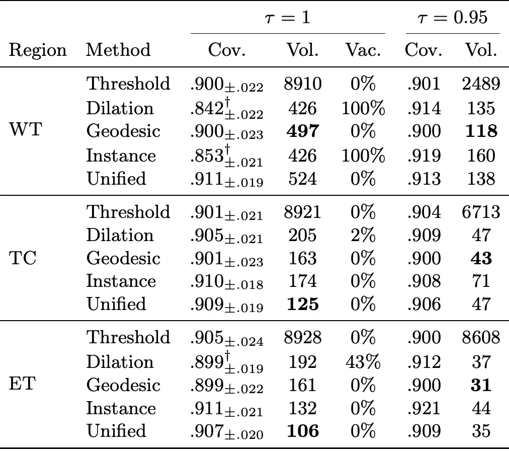
The unified method reaches joint coverage 0.911, 0.909, and 0.907 for WT, TC, and ET. Its ET volume is 45% below dilation. Instance-level dilation alone retains WT saturation; replacing its spatial geometry addresses that remaining failure.
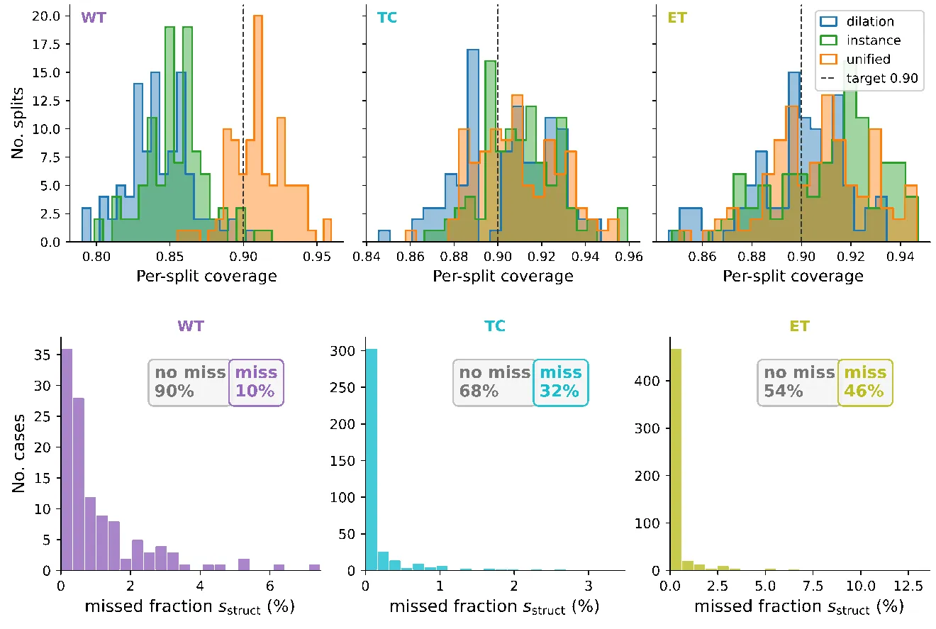
We also study conformal prediction for in-context regression, where a frozen transformer supplies the predictions used to construct response intervals.